PUBLICATIONS
Domingo-Muelas A, Duart-Abadia P, Morante-Redolat JM, Jordán-Pla A, Belenguer G, Fabra-Beser J, Paniagua-Herranz L, Pérez-Villalba A, Álvarez-Varela A, Barriga FM, Gil-Sanz C, Ortega F, Batlle E, Fariñas I. (2023). “Post-transcriptional control of a stemness signature by RNA-binding protein MEX3A regulates murine adult neurogenesis” Nat Commun. 14(1):373 https://10.1038/s41467-023-36054-6
Neural stem cells (NSCs) in the adult murine subependymal zone balance their self-renewal capacity and glial identity with the potential to generate neurons during the lifetime. Adult NSCs exhibit lineage priming via pro-neurogenic fate determinants. However, the protein levels of the neural fate determinants are not sufficient to drive direct differentiation of adult NSCs, which raises the question of how cells along the neurogenic lineage avoid different conflicting fate choices, such as self-renewal and differentiation. Here, we identify RNA-binding protein MEX3A as a post-transcriptional regulator of a set of stemness associated transcripts at critical transitions in the subependymal neurogenic lineage. MEX3A regulates a quiescence-related RNA signature in activated NSCs that is needed for their return to quiescence, playing a role in the long-term maintenance of the NSC pool. Furthermore, it is required for the repression of the same program at the onset of neuronal differentiation. Our data indicate that MEX3A is a pivotal regulator of adult murine neurogenesis acting as a translational remodeller.
Lozano-Ureña A, Lázaro-Carot L, Jiménez-Villalba E, Montalbán-Loro R, Mateos-White I, Duart-Abadía P, Martínez-Gurrea I, Nakayama KI, Fariñas I, Kirstein M, Gil-Sanz C, Ferrón SR. (2023). “IGF2 interacts with the imprinted gene Cdkn1c to promote terminal differentiation of neural stem cells.” Development. 1;150(1):dev200563 https://doi.org/10.1242/dev.200563
Adult neurogenesis is supported by multipotent neural stem cells (NSCs) with unique properties and growth requirements. Adult NSCs constitute a reversibly quiescent cell population that can be activated by extracellular signals from the microenvironment in which they reside in vivo. Although genomic imprinting plays a role in adult neurogenesis through dose regulation of some relevant signals, the roles of many imprinted genes in the process remain elusive. Insulin-like growth factor 2 (IGF2) is encoded by an imprinted gene that contributes to NSC maintenance in the adult subventricular zone through a biallelic expression in only the vascular compartment. We show here that IGF2 additionally promotes terminal differentiation of NSCs into astrocytes, neurons and oligodendrocytes by inducing the expression of the maternally expressed gene cyclin-dependent kinase inhibitor 1c (Cdkn1c), encoding the cell cycle inhibitor p57. Using intraventricular infusion of recombinant IGF2 in a conditional mutant strain with Cdkn1c-deficient NSCs, we confirm that p57 partially mediates the differentiation effects of IGF2 in NSCs and that this occurs independently of its role in cell-cycle progression, balancing the relationship between astrogliogenesis, neurogenesis and oligodendrogenesis.
Jéssica Alves Medeiros de Araújo, Soraia Barão, Isabel Mateos-White, Ana Espinosa, Marcos Romualdo Costa, Cristina Gil-Sanz, Ulrich Müller (2021). “ZBTB20 is crucial for the specification of a subset of callosal projection neurons and astrocytes in the mammalian neocortex”. Development. 148 (16): dev196642.
https://doi.org/10.1242/dev.196642
Neocortical progenitor cells generate subtypes of excitatory projection neurons in sequential order followed by the generation of astrocytes. The transcription factor zinc finger and BTB domain-containing protein 20 (ZBTB20) has been implicated in regulation of cell specification during neocortical development. Here, we show that ZBTB20 instructs the generation of a subset of callosal projections neurons in cortical layers II/III in mouse. Conditional deletion of Zbtb20 in cortical progenitors, and to a lesser degree in differentiating neurons, leads to an increase in the number of layer IV neurons at the expense of layer II/III neurons. Astrogliogenesis is also affected in the mutants with an increase in the number of a specific subset of astrocytes expressing GFAP. Astrogliogenesis is more severely disrupted by a ZBTB20 protein containing dominant mutations linked to Primrose syndrome, suggesting that ZBTB20 acts in concert with other ZBTB proteins that were also affected by the dominant-negative protein to instruct astrogliogenesis. Overall, our data suggest that ZBTB20 acts both in progenitors and in postmitotic cells to regulate cell fate specification in the mammalian neocortex.
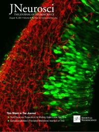
Jaime Fabra-Beser, Jessica Alves Medeiros de Araujo, Diego Marques-Coelho, Loyal A. Goff, Marcos R. Costa, Ulrich Müller, and Cristina Gil-Sanz (2021). “Differential Expression Levels of Sox9 in Early Neocortical Radial Glial Cells Regulate the Decision between Stem Cell Maintenance and Differentiation”. J. Neurosci. 41(33):6969–6986.
https://doi.org/10.1523/JNEUROSCI.2905-20.2021
Radial glial pr ogenitor cells (RGCs) in the dorsal telencephalon directly or indirectly produce excitatory projection neurons and macroglia of the neocortex. Recent evidence shows that the pool of RGCs is more heterogeneous than originally thought and that progenitor subpopulations can generate particular neuronal cell types. Using single-cell RNA sequencing, we have studied gene expression patterns of RGCs with different neurogenic behavior at early stages of cortical development. At this early age, some RGCs rapidly produce postmitotic neurons, whereas others self-renew and undergo neurogenic divisions at a later age. We have identified candidate genes that are differentially expressed among these early RGC subpopulations, including the transcription factor Sox9. Using in utero electroporation in embryonic mice of either sex, we demonstrate that elevated Sox9 expression in progenitors affects RGC cell cycle duration and leads to the generation of upper layer cortical neurons. Our data thus reveal molecular differences between progenitor cells with different neurogenic behavior at early stages of corticogenesis and indicates that Sox9 is critical for the maintenance of RGCs to regulate the generation of upper layer neurons.
de Agustín-Durán, D., Mateos-White, I., Fabra-Beser, J., and Gil-Sanz C. (2021). “Stick around: Cell-Cell Adhesion Molecules during Neocortical Development”. Cells. 10(1):118.
https://doi.org/10.3390/cells10010118
The neocortex is an exquisitely organized structure achieved through complex cellular processes from the generation of neural cells to their integration into cortical circuits after complex migration processes. During this long journey, neural cells need to establish and release adhesive interactions through cell surface receptors known as cell adhesion molecules (CAMs). Several types of CAMs have been described regulating different aspects of neurodevelopment. Whereas some of them mediate interactions with the extracellular matrix, others allow contact with additional cells. In this review, we will focus on the role of two important families of cell–cell adhesion molecules (C-CAMs), classical cadherins and nectins, as well as in their effectors, in the control of fundamental processes related with corticogenesis, with special attention in the cooperative actions among the two families of C-CAMs
Mateos-White, I., Fabra-Beser, J., de Agustín-Durán, D. and Gil-Sanz C. (2020). “Double In Utero Electroporation to Target Temporally and Spatially Separated Cell Populations”. J Vis Exp. (160).
https://doi.org/10.3791/61046
In utero electroporation is an in vivo DNA transfer technique extensively used to study the molecular and cellular mechanisms underlying mammalian corticogenesis. This procedure takes advantage of the brain ventricles to allow the introduction of DNA of interest and uses a pair of electrodes to direct the entrance of the genetic material into the cells lining the ventricle, the neural stem cells. This method allows researchers to label the desired cells and/or manipulate the expression of genes of interest in those cells. It has multiple applications, including assays targeting neuronal migration, lineage tracing, and axonal pathfinding. An important feature of this method is its temporal and regional control, allowing circumvention of potential problems related with embryonic lethality or the lack of specific CRE driver mice. Another relevant aspect of this technique is that it helps to considerably reduce the economic and temporal limitations that involve the generation of new mouse lines, which become particularly important in the study of interactions between cell types that originate in distant areas of the brain at different developmental ages. Here we describe a double electroporation strategy that enables targeting of cell populations that are spatially and temporally separated. With this approach we can label different subtypes of cells in different locations with selected fluorescent proteins to visualize them, and/or we can manipulate genes of interest expressed by these different cells at the appropriate times. This strategy enhances the potential of in utero electroporation and provides a powerful tool to study the behavior of temporally and spatially separated cell populations that migrate to establish close contacts, as well as long-range interactions through axonal projections, reducing temporal and economic costs.
Martinez-Garay, I., Gil-Sanz, C., Franco, S. J., Espinosa, A., Molnár, Z., and Mueller, U. (2016). “Cadherin 2/4 signaling via PTP1B and catenins is critical for nucleokinesis during radial neuronal migration in the neocortex”. Development 143, 2121–2134.
https://doi.org/10.1242/dev.132456
Cadherins are crucial for the radial migration of excitatory projection neurons into the developing neocortical wall. However, the specific cadherins and the signaling pathways that regulate radial migration are not well understood. Here, we show that cadherin 2 (CDH2) and CDH4 cooperate to regulate radial migration in mouse brain via the protein tyrosine phosphatase 1B (PTP1B) and α- and β-catenins. Surprisingly, perturbation of cadherin-mediated signaling does not affect the formation and extension of leading processes of migrating neocortical neurons. Instead, movement of the cell body and nucleus (nucleokinesis) is disrupted. This defect is partially rescued by overexpression of LIS1, a microtubule-associated protein that has previously been shown to regulate nucleokinesis. Taken together, our findings indicate that cadherin-mediated signaling to the cytoskeleton is crucial for nucleokinesis of neocortical projection neurons during their radial migration.
Gil-Sanz, C., Espinosa, A., Fregoso, S.P., Bluske, K.K., Cunningham, C.L., Martinez-Garay, I., Zeng, H., Franco, S.J. and Müller, U. (2015). “Lineage tracing using Cux2-Cre and Cux2-CreERT2 mice”. Neuron 86, 1091–1099.
https://doi.org/10.1016/j.neuron.2015.04.019
Using genetic fate-mapping with Cux2-Cre and Cux2-CreERT2 mice we demonstrated that the neocortical ventricular zone (VZ) contains radial glial cells (RGCs) with restricted fate potentials (Franco et al., 2012). Using the same mouse lines, Guo et al. (2013) concluded that the neocortical VZ does not contain lineage-restricted RGCs. We now show that the recombination pattern in Cux2-Cre/CreERT2 mice depends on genetic background and breeding strategies. We provide evidence that Guo et al. likely reached different conclusions because they worked with transgenic sublines with drifted transgene expression patterns. In Cux2-Cre and Cux2-CreERT2 mice that recapitulate the endogenous Cux2 expression pattern, the vast majority of fate-mapped neurons express Satb2 but not Ctip2, confirming that a restricted subset of all neocortical projection neurons belongs to the Cux2 lineage. This Matters Arising paper is in response to Guo et al. (2013), published in Neuron. See also the Matters Arising Response paper by Eckler et al. (2015), published concurrently with this Matters Arising in Neuron.
Gil-Sanz, C., Landeira, B., Ramos, C., Costa, M.R. and Müller, U. (2014). “Proliferative defects and formation of a double cortex in mice lacking Mltt4 and Cdh2 in the dorsal telencephalon”. J Neurosci 34(32):10475-87.
https://doi.org/10.1523/jneurosci.1793-14.2014
Radial glial cells (RGCs) in the ventricular neuroepithelium of the dorsal telencephalon are the progenitor cells for neocortical projection neurons and astrocytes. Here we show that the adherens junction proteins afadin and CDH2 are critical for the control of cell proliferation in the dorsal telencephalon and for the formation of its normal laminar structure. Inactivation of afadin or CDH2 in the dorsal telencephalon leads to a phenotype resembling subcortical band heterotopia, also known as «double cortex,» a brain malformation in which heterotopic gray matter is interposed between zones of white matter. Adherens junctions between RGCs are disrupted in the mutants, progenitor cells are widely dispersed throughout the developing neocortex, and their proliferation is dramatically increased. Major subtypes of neocortical projection neurons are generated, but their integration into cell layers is disrupted. Our findings suggest that defects in adherens junctions components in mice massively affects progenitor cell proliferation and leads to a double cortex-like phenotype.
Gil-Sanz, C., Franco, S.J., Martinez-Garay, I., Espinosa, A., Harkins-Perry, S. and Müller, U. (2013). “Cajal-Retzius cells instruct neuronal migration by coincidence signaling between secreted and contact-dependent guidance cues”. Neuron 79(3):461-77.
https://doi.org/10.1016/j.neuron.2013.06.040
Cajal-Retzius (CR) cells are a transient cell population of the CNS that is critical for brain development. In the neocortex, CR cells secrete reelin to instruct the radial migration of projection neurons. It has remained unexplored, however, whether CR cells provide additional molecular cues important for brain development. Here, we show that CR cells express the immunoglobulin-like adhesion molecule nectin1, whereas neocortical projection neurons express its preferred binding partner, nectin3. We demonstrate that nectin1- and nectin3-mediated interactions between CR cells and migrating neurons are critical for radial migration. Furthermore, reelin signaling to Rap1 promotes neuronal Cdh2 function via nectin3 and afadin, thus directing the broadly expressed homophilic cell adhesion molecule Cdh2 toward mediating heterotypic cell-cell interactions between neurons and CR cells. Our findings identify nectins and afadin as components of the reelin signaling pathway and demonstrate that coincidence signaling between CR cell-derived secreted and short-range guidance cues direct neuronal migration.
Franco, S.J., Gil-Sanz, C., Martinez-Garay, I., Espinosa, A., Harkins-Perry, S.R., Ramos, C. and Müller, U. (2012). “Fate-restricted neural progenitors in the mammalian cerebral cortex”. Science 337(6095):746-9.
https://doi.org/10.1126/science.1223616
During development of the mammalian cerebral cortex, radial glial cells (RGCs) generate layer-specific subtypes of excitatory neurons in a defined temporal sequence, in which lower-layer neurons are formed before upper-layer neurons. It has been proposed that neuronal subtype fate is determined by birthdate through progressive restriction of the neurogenic potential of a common RGC progenitor. Here, we demonstrate that the murine cerebral cortex contains RGC sublineages with distinct fate potentials. Using in vivo genetic fate mapping and in vitro clonal analysis, we identified an RGC lineage that is intrinsically specified to generate only upper-layer neurons, independently of niche and birthdate. Because upper cortical layers were expanded during primate evolution, amplification of this RGC pool may have facilitated human brain evolution.
Franco, S.J., Martinez-Garay, I., Gil-Sanz, C., Harkins-Perry, S.R., and Müller, U. (2011). “Reelin regulates cadherin function via Dab1/Rap1 to control neuronal migration and lamination in the neocortex”. Neuron 69(3):482-97.
https://doi.org/10.1016/j.neuron.2011.01.003
Neuronal migration is critical for establishing neocortical cell layers and migration defects can cause neurological and psychiatric diseases. Recent studies show that radially migrating neocortical neurons use glia-dependent and glia-independent modes of migration, but the signaling pathways that control different migration modes and the transitions between them are poorly defined. Here, we show that Dab1, an essential component of the reelin pathway, is required in radially migrating neurons for glia-independent somal translocation, but not for glia-guided locomotion. During migration, Dab1 acts in translocating neurons to stabilize their leading processes in a Rap1-dependent manner. Rap1, in turn, controls cadherin function to regulate somal translocation. Furthermore, cell-autonomous neuronal deficits in somal translocation are sufficient to cause severe neocortical lamination defects. Thus, we define the cellular mechanism of reelin function during radial migration, elucidate the molecular pathway downstream of Dab1 during somal translocation, and establish the importance of glia-independent motility in neocortical development.
Espinosa, A., Gil-Sanz, C., Yanagawa, Y. and Fairén A. (2009). “Two separate subtypes of early non-subplate projection neurons in the developing cerebral cortex of rodents”. Front Neuroanat. 3:27.
https://doi.org/10.3389/neuro.05.027.2009
The preplate of the cerebral cortex contains projection neurons that connect the cortical primordium with the subpallium. These are collectively named pioneer neurons. After preplate partition, most of these pioneer neurons become subplate neurons. Certain preplate neurons, however, never associate with the subplate but rather with the marginal zone. In the present overview, we propose a novel classification of non-subplate pioneer neurons in rodents into two subtypes. In rats, the neurons of the first subtype are calbindin(+) (CB), calretinin(+) (CR) and L1(+) and are situated in the upper part of the preplate before its partition. Neurons of the second subtype are TAG-1(+) and are located slightly deeper to the previous population in the preplate. After the preplate partition, the CB(+), CR(+) and L1(+) neurons remain in the marginal zone whereas TAG-1(+) neurons become transiently localized in the upper cortical plate. In mice, by contrast, calcium binding proteins did not label pioneer neurons. We define in mice two subtypes of non-subplate pioneer neurons, either L1(+) or TAG-1(+)/cntn2(+). We propose these to be the homologues of the two subtypes of non-subplate pioneer neurons of rats. The anatomical distribution of these neuron populations is similar in rats and mice. The two populations of non-subplate pioneer neurons differ in their axonal projections. Axons of L1(+) pioneer neurons project to the ganglionic eminences and the anterior preoptic area, but avoid entering the posterior limb of the internal capsule towards the thalamus. Axons of TAG-1(+) pioneer neurons project to the lateral parts of the ganglionic eminences at the early stages of cortical histogenesis examined.
